Transkript
Anmerkung:
Es folgt eine detaillierte Auflistung der Literatur, die für dieses Video als inhaltliche Grundlage und/oder Bildvorlage genutzt wurde. Außerdem wird hier um weiterführende Literatur ergänzt, die bestimmte Aspekte vertieft. Es gibt keinen Anspruch auf Vollständigkeit. Die Literatur ist nicht zwingend alphabetisch geordnet, eher nach Wichtigkeit innerhalb eines Kapitels. Hinter einigen Literaturquellen stehen in eckigen Klammern einige Kommentare meinerseits. Ggfs. wird die Liste aktualisiert.
Domänen des Lebens
Es gibt eine Reihe an Standardliteratur, die sich mit den Domänen des Lebens befasst. Für eine weiterführende Literaturrecherche lohnt sich ein Blick in die Literaturverzeichnisse dieser Lehrbücher, da dort viele Primärpublikationen gelistet sind.
- Madigan, M.T., et al.: Brock Biology of Microorganisms
(aktuelle Auflagen, z.B. 16. Auflage, 2021)
- Rosenberg et al. (Eds): The Prokaryotes: Prokaryotic Biology and Symbiotic Associations (Springer, mehrere Bände, aktuelle Auflagen)
- Campbell, N.A., Reece, J.B. et al: Biology (z.B. 12. Auflage, 2020); Kapitel zur Systematik und Evolution der Domänen.
- Alberts, B. et al.: Molecular Biology of the Cell (aktuelle Auflagen, z.B. 7. Auflage, 2022); Abschnitte zu Unterschieden zwischen Prokaryoten und Eukaryoten.
- Futuyma, D.J., Kirkpatrick, M.: Evolution (z.B. 5. Auflage, 2023), Systematik, phylogenetische Einordnung und Evolution der Domänen.
- Mein Artikel „Entstehung des Lebens Kapitel 10: Last Universal Common Ancestor (LUCA)“ befasst sich ebenfalls mit den Domänen des Lebens und ihrer phylogenetischen Beziehungen: https://internet-evoluzzer.de/last-universal-common-ancestor-luca/
Klassische Arbeiten & Reviews zur Domäneneinteilung:
- Woese, C.R., Kandler, O., Wheelis, M.L. (1990): „Towards a natural system of organisms: Proposal for the domains Archaea, Bacteria, and Eucarya.“ Proceedings of the National Academy of Sciences 87(12): 4576–4579. [Originalarbeit, die die Dreiteilung vorschlägt.]
- Hug, L.A., et al. (2016): „A new view of the tree of life.“ Nature Microbiology 1, 16048. [Aktuelle phylogenomische Sicht, viele neue prokaryotische Linien.]
- Williams, T.A., et al. (2021): „Phylogenomics provides robust support for a two-domains tree of life.“ Nature Ecology & Evolution 5: 123–133. [Diskussion über die Zwei- vs. Drei-Domänen-Hypothese.]
- Cavalier-Smith, T. (2002): „The neomuran origin of archaebacteria, the negibacterial root of the universal tree and bacterial megaclassification.“ International Journal of Systematic and Evolutionary Microbiology 52(1): 7–76. [Alternative Hypothesen und Megaklassifikation.]
Vielfalt der Bakterien:
- Dykhuizen, Daniel. (2005). Species Numbers in Bacteria. Proceedings. California Academy of Sciences. 56. 62-71. [Artenzahlen der Bakterien]
- Zhu, Daochen & Hosoi-Tanabe, Shoko & Yang, Chong & Zhang, Weimin & Sun, Jianzhong. (2013). Bacterial Community Composition of South China Sea Sediments through Pyrosequencing-Based Analysis of 16S rRNA Genes. PloS one. 8. e78501. 10.1371/journal.pone.0078501. [Artenvielfalt der Bakterien]
- Bertin, P., Heinrich-Salmeron, A., Pelletier, E. et al. Metabolic diversity among main microorganisms inside an arsenic-rich ecosystem revealed by meta- and proteo-genomics. ISME J 5, 1735–1747 (2011). https://doi.org/10.1038/ismej.2011.51 [Artenvielfalt der Bakterien]
- Parks, D.H., Rinke, C., Chuvochina, M. et al. Recovery of nearly 8,000 metagenome-assembled genomes substantially expands the tree of life. Nat Microbiol 2, 1533–1542 (2017). https://doi.org/10.1038/s41564-017-0012-7 [Artenvielfalt der Bakterien]
- Anantharaman, K., Brown, C., Hug, L. et al. Thousands of microbial genomes shed light on interconnected biogeochemical processes in an aquifer system. Nat Commun 7, 13219 (2016). https://doi.org/10.1038/ncomms13219 [Artenvielfalt der Bakterien]
- Parte, A. C. et al. (2020): List of Prokaryotic names with Standing in Nomenclature (LPSN) moves to the DSMZ. International Journal of Systematic and Evolutionary Microbiology. https://doi.org/10.1099/ijsem.0.004332 [Bakterien-Phyla]
- Oren, A., Garrity, GM. (2021): Valid publication of the names of forty-two phyla of prokaryotes. International Journal of Systematic and Evolutionary Microbiology 71. https://doi.org/10.1099/ijsem.0.005056 [Bakterien-Phyla]
- Dudek, Natasha K. et al. (2017): Novel Microbial Diversity and Functional Potential in the Marine Mammal Oral Microbiome. Current Biology, Volume 27, Issue 24, 3752 – 3762.e6 [Vielfalt der Bakterien]
- Brendan B. Larsen, Elizabeth C. Miller, Matthew K. Rhodes, and John J. Wiens: Inordinate Fondness Multiplied and Redistributed: the Number of Species on Earth and the New Pie of Life. The Quarterly Review of Biology 2017 92:3, 229-265 [potentielle Artenvielfalt der Bakterien]
- Pallen, Mark & Telatin, Andrea & Oren, Aharon. (2020). The Next Million Names for Archaea and Bacteria. Trends in Microbiology. 29. 10.1016/j.tim.2020.10.009. [potentielle Artenvielfalt der Bakterien und Archaeen]
- J. Locey,& J.T. Lennon, Scaling laws predict global microbial diversity, Proc. Natl. Acad. Sci. U.S.A. 113 (21) 5970-5975, https://doi.org/10.1073/pnas.1521291113 (2016). [potentielle Artenvielfalt der Bakterien und Archaeen]
- Ferla MP, Thrash JC, Giovannoni SJ, Patrick WM. New rRNA gene-based phylogenies of the Alphaproteobacteria provide perspective on major groups, mitochondrial ancestry and phylogenetic instability. PLoS One. 2013 Dec 11;8(12):e83383. doi: 10.1371/journal.pone.0083383. PMID: 24349502; PMCID: PMC3859672. [Alphaproteobacteria]
- Rizzatti G, Lopetuso LR, Gibiino G, Binda C, Gasbarrini A (2017). „Proteobacteria: A Common Factor in Human Diseases“. BioMed Research International. 2017: 9351507. doi:10.1155/2017/9351507. PMC 5688358. PMID 29230419. [Alphaproteobacteria]
- Kersters K, De Vos P, Gillis M, Swings J, Vandamme P, Stackebrandt E (2006). „Introduction to the Proteobacteria“. In Dworkin M, Falkow S, Rosenberg E, Schleifer KH (eds.). The Prokaryotes. Vol. 5: Proteobacteria: Alpha and Beta Subclasses. New York, NY: Springer. pp. 3–37. doi:10.1007/0-387-30745-1_1. ISBN 978-0-387-30745-9. [Alphaproteobacteria]
- Whitton, Brian A.; Potts, Malcolm, eds. (2002). The Ecology of Cyanobacteria. doi:10.1007/0-306-46855-7. [Cyanobacteria]
- Oren, Aharon; Mareš, Jan; Rippka†, Rosmarie (2022). „Validation of the names Cyanobacterium and Cyanobacterium stanieri, and proposal of Cyanobacteriota phyl. nov“. International Journal of Systematic and Evolutionary Microbiology. 72 (10): 005528 [Cyanobactiera]
- Migula W (1895). „Bacteriaceae (Stabchenbacterien)“. In Engerl A, Prantl K (eds.). Die Naturlichen Pfanzenfamilien, W. Engelmann, Leipzig, Teil I, Abteilung Ia. pp. 20–30. [Coli-Bakterien]
- Castellani A, Chalmers AJ (1919). Manual of Tropical Medicine (3rd ed.). New York: Williams Wood and Co [Coli-Bakterien]
Vielfalt der Archaeen:
- Williams TA, Szöllősi GJ, Spang A, Foster PG, Heaps SE, Boussau B, et al. (2017). „Integrative modeling of gene and genome evolution roots the archaeal tree of life“. Proceedings of the National Academy of Sciences of the United States of America. 114 (23): E4602 – E4611. doi:10.1073/pnas.1618463114.
- Poli, Annarita & Finore, Ilaria & Romano, Ida & Gioiello, Alessia & Lama, Licia & Nicolaus, Barbara. (2017). Microbial Diversity in Extreme Marine Habitats and Their Biomolecules. Microorganisms. 5. 10.3390/microorganisms5020025.
- Castelle, Cindy J. et al. (2015): Genomic Expansion of Domain Archaea Highlights Roles for Organisms from New Phyla in Anaerobic Carbon Cycling Current Biology, Volume 25, Issue 6, 690 – 701
- Castelle CJ, Banfield JF (2018). „Major New Microbial Groups Expand Diversity and Alter our Understanding of the Tree of Life“. Cell. 172 (6): 1181–1197.
- Magnuson, E., Altshuler, I., Fernández-Martínez, M.Á. et al. Active lithoautotrophic and methane-oxidizing microbial community in an anoxic, sub-zero, and hypersaline High Arctic spring. ISME J 16, 1798–1808 (2022). https://doi.org/10.1038/s41396-022-01233-8 [mikrobiellen Gemeinschaft im Lost Hammer Spring]
Bakterien bzw. Mikroben auf bzw. im menschlichen Körper:
- Sender, Ron; Fuchs, Shai; Milo, Ron (2016). „Revised Estimates for the Number of Human and Bacteria Cells in the Body“. PLOS Biology. 14 (8): e1002533. doi:10.1371/journal.pbio.1002533.
- Lacy, Brian E.; Spiegel, Brennan (2019). „Introduction to the Gut Microbiome Special Issue“. American Journal of Gastroenterology. 114 (7): 1013. doi:10.14309/ajg.0000000000000303.
- Sung, Jaeyun; et al. (2024). „The human gut microbiome in critical illness: disruptions, consequences, and therapeutic frontiers“. Journal of Critical Care. 79: 154436.
Domäne der Eukaryoten:
- Keeling PJ, Eglit Y. Openly available illustrations as tools to describe eukaryotic microbial diversity. PLoS Biol. 2023 Nov 21;21(11):e3002395. doi: 10.1371/journal.pbio.3002395. PMID: 37988341; PMCID: PMC10662721.
- Burki F (2014). „The eukaryotic tree of life from a global phylogenomic perspective“. Cold Spring Harbor Perspectives in Biology. 6 (5): a016147. doi:10.1101/cshperspect.a016147
- Burki F, Kaplan M, Tikhonenkov DV, et al. (2016). „Untangling the early diversification of eukaryotes: a phylogenomic study of the evolutionary origins of Centrohelida, Haptophyta and Cryptista“. Proceedings: Biological Sciences. 283 (1823): 20152802. doi:10.1098/rspb.2015.2802.
- Adl SM, Bass D, Lane CE, et al. (January 2019). „Revisions to the Classification, Nomenclature, and Diversity of Eukaryotes“. The Journal of Eukaryotic Microbiology. 66 (1): 4–119. doi:10.1111/jeu.12691.
- Brown MW, Heiss AA, Kamikawa R, Inagaki Y, Yabuki A, Tice AK, Shiratori T, Ishida KI, Hashimoto T, Simpson A, Roger A (19 January 2018). „Phylogenomics Places Orphan Protistan Lineages in a Novel Eukaryotic Super-Group“. Genome Biology and Evolution. 10 (2): 427–433. doi:10.1093/gbe/evy014.
- Margulis, L., Chapman, M.J. (2009): „Kingdoms and Domains: An Illustrated Guide to the Phyla of Life on Earth.“ Elsevier. [Ein klassisches Werk mit Fokus auf morphologische Vielfalt.]
- Brown, M.W., Heiss, A.A., Kamikawa, R., et al. (2018): „Phylogenomics places orphan protistan lineages in a novel eukaryotic super-group.“ Genome Biology and Evolution 10(2): 427–433.
- Derelle, R., Torruella, G., Klimeš, V., et al. (2015): „Bacterial proteins pinpoint a single eukaryotic root.“ Proceedings of the National Academy of Sciences 112(7): E693–E699.
- Butterfield, N.J. (2015). „Early evolution of the Eukaryota“. Palaeontology. 58 (1): 5–17
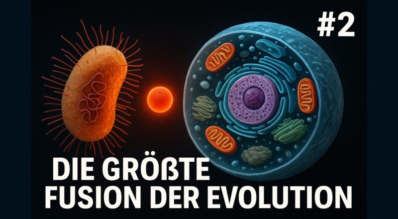
Endosymbiontentheorie
Klassische, historische und allgemeine Literatur zur Endosymbiontentheorie:
- Lynn Margulis (1967): „On the origin of mitosing cells.“ Journal of Theoretical Biology 14(3): 225–274. [Ursprüngliche Formulierung der Endosymbiontentheorie für Mitochondrien.]
- Lynn Margulis: Origin of Eukaryotic Cells. Yale University Press, New Haven 1970.
- Lynn Margulis, Dorion Sagan: Microcosmos: Four Billion Years of Microbial Evolution, University of California Press, Berkeley, 1997, ISBN 978-0520210646
- Margulis, Lynn; Chapman, Michael; Guerrero, Ricardo; Hall, John (2006). „The last eukaryotic common ancestor (LECA): Acquisition of cytoskeletal motility from aerotolerant spirochetes in the Proterozoic Eon“. Proceedings of the National Academy of Sciences. 103 (35): 13080–13085.
- Mereschkowski, K. S. : Über Natur und Ursprung der Chromatophoren im Pflanzenreiche. In: Biologisches Centralblatt. Band 25, 15. September 1905, S. 593–604 (englisch)
- Maynard Smith J, Szathmary E. The Major Transitions in Evolution. Oxford University Press, Oxford (1995).
- Lane, N. (2009): Life Ascending: The Ten Great Inventions of Evolution. WW Norton/Profile, London.
- Lane, N. (2017): Der Funke des Lebens Energie und Evolution. Konrad Theis Verlag
- Pisani D, Cotton JA, McInerney JO. Supertrees disentangle the chimeric origin of eukaryotic genomes. Molecular Biology Evolution 24: 1752–60 (2007).
- Archibald, J.M. (2015): „Endosymbiosis and Eukaryotic Cell Evolution.“ Current Biology 25(19): R911–R921. [Prägnanter Überblick über primäre, sekundäre und tertiäre Endosymbiosen.]
- Roger, A.J., Muñoz-Gómez, S.A., Kamikawa, R. (2017): „The Origin and Diversification of Mitochondria.“ Current Biology 27(21): R1177–R1192. [Beleuchtet mitochondriale Ursprünge im Kontext der eukaryotischen Diversifizierung.]
- Nowack, E.C.M., Weber, A.P.M. (2018): „Genomics-informed insights into endosymbiotic organelle evolution in photosynthetic eukaryotes.“ Annual Review of Plant Biology 69: 51–84. [Review über Plastidenentstehung und Endosymbiose in Algen.]
- Martin, W.F., Garg, S., Zimorski, V. (2015): „Endosymbiotic theories for eukaryote origin.“ Philosophical Transactions of the Royal Society B 370(1678): 20140330. [Verteidigung der Symbiogenese als zentrale Erklärung.]
- Latorre A, Durban A, Moya A, Pereto J (2011). „The role of symbiosis in eukaryotic evolution“. In Gargaud M, López-Garcìa P, Martin H (eds.). Origins and Evolution of Life: An astrobiological perspective. Cambridge: Cambridge University Press. pp. 326–339.
- Gabaldón T (October 2021). „Origin and Early Evolution of the Eukaryotic Cell“. Annual Review of Microbiology. 75 (1): 631–647.
- O’Malley MA, Leger MM, Wideman JG, Ruiz-Trillo I (2019). „Concepts of the last eukaryotic common ancestor“. Nature Ecology & Evolution. 3 (3): 338–344.
- Strassert, Jürgen F. H.; Irisarri, Iker; Williams, Tom A.; Burki, Fabien (2021). „A molecular timescale for eukaryote evolution with implications for the origin of red algal-derived plastids“. Nature Communications. 12 (1): 1879.
- Leander BS (2020). „Predatory protists“. Current Biology. 30 (10): R510 – R516
- Bremer, Nico; Tria, Fernando D. K.; Skejo, Josip; Garg, Sriram G.; Martin, William F. (2022). „Ancestral State Reconstructions Trace Mitochondria But Not Phagocytosis to the Last Eukaryotic Common Ancestor“. Genome Biology and Evolution. 14 (6).
- Makarova, Kira S.; Yutin, Natalya; Bell, Stephen D.; Koonin, Eugene V. (2010). „Evolution of diverse cell division and vesicle formation systems in Archaea“. Nature Reviews Microbiology. 8 (10): 731–741.
Aktuelle Endosymbioseforschung (Nitroblasten):
- Wong, Carissa (2024). „Scientists discover first algae that can fix nitrogen — thanks to a tiny cell structure“. Nature. 628 (8009): 702.
- H. Coale et al. Nitrogen-fixing organelle in a marine alga. Science. Vol. 384, April 12, 2024, p. 217. doi: 10.1126/science.adk1075.
Entstehung der Eukaryoten, gerade auch im Zusammenhang der Asgard-Archaeen:
- Eme, L., Spang, A., Lombard, J., Stairs, C.W., Ettema, T.J.G. (2017): „Archaea and the origin of eukaryotes.“ Nature Reviews Microbiology 15, 711–723. [Review zu Archaeen, Asgard-Archaeen und Eukaryoten.]
- Spang, A., Saw, J.H., Jørgensen, S.L., et al. (2015): „Complex archaea that bridge the gap between prokaryotes and eukaryotes.“ Nature 521, 173–179. [Asgard-Archaea, Bedeutung für Eukaryotenentstehung.]
- Imachi, H., et al. (2020): „Isolation of an archaeon at the prokaryote–eukaryote interface.“ Nature 577, 519–525. [Lebendnachweis von Lokiarchaeota, relevant für Ursprung der Eukaryoten.]
- Koonin, E.V. (2015): „The Origin and Early Evolution of Eukaryotes in the Light of Phylogenomics.“ Genome Biology 16, 1–17.
- Koonin, E.V., Yutin, N. (2014): „The Dispersed Archaeal Eukaryome: Roots of Eukaryotic Complexity.“ Current Opinion in Microbiology 22: 67–74.
- Koonin, Eugene V. (March 2005). „The incredible expanding ancestor of eukaryotes“. Cell. 140 (5): 606–608.
- Williams, Tom A.; Foster, Peter G.; Cox, Cymon J.; Embley, T. Martin (2013). An archaeal origin of eukaryotes supports only two primary domains of life. Nature, 504(7479), 231–236. doi:10.1038/nature12779
- Liao Y, Ithurbide S, de Silva RT, Erdmann S, Duggin IG. Archaeal cell biology: diverse functions of tubulin-like cytoskeletal proteins at the cell envelope. Emerg Top Life Sci. 2018 Dec 14;2(4):547-559. doi: 10.1042/ETLS20180026. PMID: 33525829.
- Felipe MerinoStefan Raunser (2016) Cytoskeleton: The mother of all actins? eLife 5:e23354.
- Stairs CW, Ettema TJG. The Archaeal Roots of the Eukaryotic Dynamic Actin Cytoskeleton. Curr Biol. 2020 May 18;30(10):R521-R526. doi: 10.1016/j.cub.2020.02.074. PMID: 32428493.
- Caner Akıl, Linh T. Tran, Magali Orhant-Prioux, Yohendran Baskaran, Edward Manser, Laurent Blanchoin, Robert C. Robinson. Complex eukaryotic-like actin regulation systems from Asgard archaea. bioRxiv 768580; doi: https://doi.org/10.1101/768580
- Tarrason Risa G, Hurtig F, Bray S, Hafner AE, Harker-Kirschneck L, Faull P, Davis C, Papatziamou D, Mutavchiev DR, Fan C, Meneguello L, Arashiro Pulschen A, Dey G, Culley S, Kilkenny M, Souza DP, Pellegrini L, de Bruin RAM, Henriques R, Snijders AP, Šarić A, Lindås AC, Robinson NP, Baum B. The proteasome controls ESCRT-III-mediated cell division in an archaeon. Science. 2020 Aug 7;369(6504):eaaz2532. doi: 10.1126/science.aaz2532.
- Lu ZFu TLi TLiu Y, Zhang SLi J, Dai JKoonin EV, Li G, Chu H, Li M. 2020. Coevolution of Eukaryote-like Vps4 and ESCRT-III Subunits in the Asgard Archaea. mBio 11:10.1128/mbio.00417-20. https://doi.org/10.1128/mbio.00417-20
- C. Tromer,J.J.E. van Hooff,G.J.P.L. Kops,& B. Snel, Mosaic origin of the eukaryotic kinetochore, Proc. Natl. Acad. Sci. U.S.A. 116 (26) 12873-12882, https://doi.org/10.1073/pnas.1821945116 (2019).
- Rodrigues-Oliveira, T., Wollweber, F., Ponce-Toledo, R.I. et al. Actin cytoskeleton and complex cell architecture in an Asgard archaeon. Nature 613, 332–339 (2023). https://doi.org/10.1038/s41586-022-05550-y
- Charles-Orszag A, Petek-Seoane NA, Mullins RD. 2024. Archaeal actins and the origin of a multi-functional cytoskeleton. J Bacteriol 206:e00348-23. https://doi.org/10.1128/jb.00348-23
- Williams TA, Foster PG, Cox CJ, Embley TM. An archaeal origin of eukaryotes supports only two primary domains of life. Nature 504: 231–36 (2013).
Introns und die Entstehung des Zellkerns
Guter Überblicksartikel zur Entstehung des Zellkerns:
- Martin WF, Garg S, Zimorski V. Endosymbiotic theories for eukaryote origin. Philos Trans R Soc Lond B Biol Sci. 2015 Sep 26;370(1678):20140330. doi: 10.1098/rstb.2014.0330. PMID: 26323761; PMCID: PMC4571569.
Aktuelle Reviews zur Evolution des Zellkerns
- Alberts, B. et al. (2022): „Molecular Biology of the Cell“ (7th edition). Kapitel zur Nuclear Envelope and Traffic Between the Nucleus and Cytoplasm. [Molekulare Grundlagen der Kernstruktur mit evolutionären Aspekten.]
- Mans, B.J., Anantharaman, V., Aravind, L., Koonin, E.V. (2004): „Comparative genomics, evolution and origins of the nuclear envelope and nuclear pore complex.“ Cell Cycle 3(12): 1612–1637. [Klassischer Review zu Komponenten des Zellkerns aus genomischer Sicht.]
- Field, M.C., Rout, M.P. (2007): „Pore timing: the evolutionary origins of the nucleus and nuclear pore complex.“ FEMS Microbiology Reviews 31(3): 345–351. [Überblick über die Evolution der Kernporenkomplexe.]
- Neumann, N., Lundin, D., Poole, A.M. (2010): „Comparative genomic evidence for a complete nuclear pore complex in the last eukaryotic common ancestor.“ PLoS ONE 5(10): e13241. [Belege für die frühe Evolution des Kernporenapparats.]
- Dacks, J.B., Devos, D.P. (2021): „The origin and evolution of the eukaryotic endomembrane system.“ Current Biology 31(10): R497–R510. [Erweiterter Kontext des Zellkerns im endomembranären System.]
Literatur zu Gemmata obscuriglobus
- Devos, Damien P. (2013). Gemmata obscuriglobus. Current Biology, Volume 23, Issue 17, R705 – R707
- Santarella-Mellwig R, Franke J, Jaedicke A, Gorjanacz M, Bauer U, Budd A, et al. (2010) The Compartmentalized Bacteria of the Planctomycetes-Verrucomicrobia-Chlamydiae Superphylum Have Membrane Coat-Like Proteins. PLoS Biol 8(1): e1000281. https://doi.org/10.1371/journal.pbio.1000281
Introns, Exons und alternatives Spleißen:
- Martin, William; Koonin, Eugene V. (2006). „Introns and the origin of nucleus–cytosol compartmentalization“. Nature. 440 (7080): 41–45. [Rolle der Introns im Zusammenhang der Entstehung des Zellkerns]
- Koonin, E. V. (2006). The origin of introns and their role in eukaryogenesis: a compromise solution to the introns-early versus introns-late debate? Biology Direct, 1(1), 22. DOI: 10.1186/1745-6150-1-22
- Timmis, J., Ayliffe, M., Huang, C. et al. Endosymbiotic gene transfer: organelle genomes forge eukaryotic chromosomes. Nat Rev Genet 5, 123–135 (2004). https://doi.org/10.1038/nrg1271
- Lane, Nick; Martin, William F. (2010). „The energetics of genome complexity“. Nature. 467 (7318): 929–934
- Lambowitz AM, Zimmerly S. Group II introns: mobile ribozymes that invade DNA. Cold Spring Harb Perspect Biol. 2011 Aug 1;3(8):a003616. doi: 10.1101/cshperspect.a003616. PMID: 20463000; PMCID: PMC3140690.
- Roitzsch, M., Fedorova, O. & Pyle, A. The 2′-OH group at the group II intron terminus acts as a proton shuttle. Nat Chem Biol 6, 218–224 (2010). https://doi.org/10.1038/nchembio.312
- Irimia, M., & Roy, S. W. (2014). Origin of spliceosomal introns and alternative splicing. Cold Spring Harbor Perspectives in Biology, 6(6), a016071. DOI: 10.1101/cshperspect.a016071 [Umfassender Überblick über die Entstehung von Introns und die Evolution des alternativen Spleißens].
- Roy, S. W., & Gilbert, W. (2006). The evolution of spliceosomal introns: patterns, puzzles and progress. Nature Reviews Genetics, 7(3), 211–221. DOI: 10.1038/nrg1807
- Keren, H., Lev-Maor, G., & Ast, G. (2010). Alternative splicing and evolution: diversification, exon definition and function. Nature Reviews Genetics, 11(5), 345–355. DOI: 10.1038/nrg2776 [Verbindet alternative Spleißmechanismen mit der evolutionären Diversität]
- Alberts, B. et al.: Molecular Biology of the Cell (aktuelle Auflagen, z.B. 7. Auflage, 2022); Kapitel: RNA Processing and Posttranscriptional Control. [Enthält Grundlagen zum Splicing und seinen evolutionären Implikationen]
- Morgan JT, Fink GR, Bartel DP. Excised linear introns regulate growth in yeast. Nature. 2019 Jan;565(7741):606-611. doi: 10.1038/s41586-018-0828-1. Epub 2019 Jan 16. PMID: 30651636; PMCID: PMC6464110.
- Tan JH, Fraser AG. The combinatorial control of alternative splicing in C. elegans. PLoS Genet. 2017 Nov 9;13(11):e1007033. doi: 10.1371/journal.pgen.1007033. PMID: 29121637; PMCID: PMC5697891.
- Celotto AM, Graveley BR. Alternative splicing of the Drosophila Dscam pre-mRNA is both temporally and spatially regulated. Genetics. 2001 Oct;159(2):599-608. doi: 10.1093/genetics/159.2.599. PMID: 11606537; PMCID: PMC1461822.
- Chorev M, Carmel L. The function of introns. Front Genet. 2012 Apr 13;3:55. doi: 10.3389/fgene.2012.00055. PMID: 22518112; PMCID: PMC3325483.
- Lane N. Plastids, genomes and the probability of gene transfer. Genome Biology and Evolution 3: 372–74 (2011).
- Lane N. Power, Sex, Suicide: Mitochondria and the Meaning of Life. Oxford University Press, Oxford.
- Lane, N. (2009): Life Ascending: The Ten Great Inventions of Evolution. WW Norton/Profile, London.
- Lane, N. (2017): Der Funke des Lebens Energie und Evolution. Konrad Theis Verlag
Entgegnung der kreationistischen Kritik an der Endosymbiontentheorie:
- Die Grundlagen der Endosymbiontentheorie (AG Evolutionsbiologie) https://www.ag-evolutionsbiologie.de/html/2022/endosymbiontentheorie-evolution.html
Komplexe Genome
Histone:
- Alberts, B. et al.: Molecular Biology of the Cell (aktuelle Auflagen, z.B. 7. Auflage, 2022) Kapitel: Chromatin Structure and Function [Behandelt neben dem allgemeinen Aufbau auch evolutionäre Aspekte und funktionelle Divergenz der Histonvarianten.]
- Pontremoli, C., et al. (2021). Universal and taxon-specific trends in protein sequences as a function of age. eLife, 10, e59391. DOI: 10.7554/eLife.59391 [Histone gehören zu den evolutionär ältesten Proteinen – Diskussion ihrer Ursprünge im LUCA-Kontext.]
- Henneman B, van Emmerik C, van Ingen H, Dame RT (2018). „Structure and function of archaeal histones“. PLOS Genetics. 14 (9): e1007582.
- Rizzo PJ (August 2003). „Those amazing dinoflagellate chromosomes“. Cell Research. 13 (4): 215–7. doi:10.1038/sj.cr.7290166
- Talbert PB, Henikoff S (December 2012). „Chromatin: packaging without nucleosomes“. Current Biology. 22 (24): R1040-3. Bibcode:2012CBio…22R1040T. doi:10.1016/j.cub.2012.10.052
- Lei, Bingkun & Capella, Matías & Montgomery, Sean & Borg, Michael & Osakabe, Akihisa & Goiser, Malgorzata & Muhammad, Abubakar & Braun, Sigurd & Berger, Frederic. (2020). A Synthetic Approach to Reconstruct the Evolutionary and Functional Innovations of the Plant Histone Variant H2A.W. Current Biology. 31. 10.1016/j.cub.2020.09.080.
Populärwissenschaftliche Literatur zur Epigenetik:
- Nessa Carey (2012). The Epigenetics Revolution: How Modern Biology Is Rewriting Our Understanding of Genetics, Disease and Inheritance [Eines der bekanntesten Bücher über Epigenetik. Klar und spannend erklärt – von Grundlagen bis zu Auswirkungen auf Gesundheit, Evolution und Gesellschaft.]
- Eva Jablonka & Marion J. Lamb (2014). Evolution in Four Dimensions: Genetic, Epigenetic, Behavioral, and Symbolic Variation in the History of Life [Verbindet Epigenetik mit Evolutionstheorie.]
Histonmodifikationen:
- G. Goll, T. H. Bestor: Histone modification and replacement in chromatin activation. Genes Dev 16(14): S. 1739-1742 (2002) PMID 12130533
- A. Grant: A tale of histone modifications. Genome Biol 2(4):REVIEWS0003 (2001)
- J. Bannister, R. Schneider, T. Kouzarides: Histone methylation: dynamic or static? Cell 109(7): S. 801-806 (2002) PMID 12110177
- Cheung, C. D. Allis, P. Sassone-Corsi: Signaling to chromatin through histone modifications. Cell 103(2): S. 263-271 (2000) PMID 11057899
- L. Peterson, M. A. Laniel: Histones and histone modifications. Curr Biol 14(14): S. R546-51 (2004) PMID 15268870
- Shi, J. R. Whetstine: Dynamic regulation of histone lysine methylation by demethylases. Mol Cell 25(1): S. 1-14 (2007) PMID 17218267
- Wang, Z., Zang, C., Rosenfeld, J. et al. Combinatorial patterns of histone acetylations and methylations in the human genome. Nat Genet 40, 897–903 (2008). https://doi.org/10.1038/ng.154
- Jenuwein, T., & Allis, C. D. (2001). Translating the histone code. Science, 293(5532), 1074–1080. DOI: 10.1126/science.1063127 [Bedeutung der posttranslationalen Modifikationen in der Evolution epigenetischer Regulation.]
Allgemeine populärwissenschaftliche Lehrbücher Transkriptionsfaktoren und Genregulation und ihrer Rolle in der Evolution:
- Alberts, B. et al.: Molecular Biology of the Cell (aktuelle Auflagen, z.B. 7. Auflage, 2022) Kapitel: „Control of Gene Expression“, „Transcription Regulators in Eukaryotes“, „Chromatin Remodeling“, „Development and Differentiation“
- David Latchman – Gene Regulation: A Eukaryotic Perspective 7th Edition (2021). Fokus: Komplettes Werk zur eukaryotischen Genregulation – Schwerpunkt Transkriptionsfaktoren
- Davidson, E. H. – The Regulatory Genome: Gene Regulatory Networks in Development and Evolution (2006). [Fachbuch mit starker Betonung auf GRNs und deren evolutionärer Bedeutung]
- Carroll, S. B. – Endless Forms Most Beautiful: The New Science of Evo Devo. [Populärwissenschaftlich mit Tiefgang, Fokus: Evolution der Genregulation als Triebkraft der morphologischen Vielfalt, Besonderheit: Hervorragende Beispiele zu Hox-Genen, cis-regulatorischen Elementen, TF-Netzwerken; auch in deutscher Sprache unter dem Titel „EVO DEVO: Das neue Bild der Evolution“
Fachartikel zur Evolution der Transkriptionsfaktoren
- Levine, M., & Tjian, R. (2003). Transcription regulation and animal diversity. Nature, 424(6945), 147–151. DOI: 10.1038/nature01763 [Schlüsselarbeit zur Rolle von TFs bei der Entstehung tierischer Vielfalt.]
- Wray, G. A. (2007). The evolutionary significance of cis-regulatory mutations. Nature Reviews Genetics, 8(3), 206–216. DOI: 10.1038/nrg2063 [Diskussion zur Wechselwirkung von TFs und cis-regulatorischen Elementen.]
- Minezaki Y, Homma K, Kinjo AR, Nishikawa K. Human transcription factors contain a high fraction of intrinsically disordered regions essential for transcriptional regulation. J Mol Biol. 2006 Jun 16;359(4):1137-49. doi: 10.1016/j.jmb.2006.04.016. Epub 2006 Apr 25. Erratum in: J Mol Biol. 2006 Oct 13;363(1):309. PMID: 16697407. [Literatur über die intristically disordered regions bei Transkriptionsfaktoren, die im Video angesprochen wurden]
- Long, H. K., Prescott, S. L., & Wysocka, J. (2016). Ever-Changing Landscapes: Transcriptional Enhancers in Development and Evolution. Cell, 167(5), 1170–1187. DOI: 10.1016/j.cell.2016.09.018
- Rebeiz, M., & Tsiantis, M. (2017). Enhancer evolution and the origins of morphological novelty. Current Opinion in Genetics & Development, 45, 115–123. DOI: 10.1016/j.gde.2017.04.006
- Erwin, D. H., & Davidson, E. H. (2009). The evolution of hierarchical gene regulatory networks. Science, 324(5923), 1121–1125. DOI: 10.1126/science.1171651
- Peter, I. S., & Davidson, E. H. (2011). Evolution of gene regulatory networks controlling body plan development. Cell, 144(6), 970–985. DOI: 10.1016/j.cell.2011.02.017
- True, J. R., & Carroll, S. B. (2002). Gene co-option in physiological and morphological evolution. Annual Review of Cell and Developmental Biology, 18, 53–80. DOI: 10.1146/annurev.cellbio.18.020402.140619
- Tuch, B. B., Li, H., & Johnson, A. D. (2008). Evolution of eukaryotic transcription circuits. Science, 319(5871), 1797–1799. DOI: 10.1126/science.1152398
- de Mendoza, A., Sebé-Pedrós, A., & Ruiz-Trillo, I. (2013). Transcription factor evolution in eukaryotes and the assembly of the regulatory toolkit in multicellular lineages. Proceedings of the National Academy of Sciences, 110(50), E4858–E4866. DOI: 10.1073/pnas.1311818110
- McGinnis, W., & Krumlauf, R. (1992). Homeobox genes and axial patterning. Cell, 68(2), 283–302. DOI: 10.1016/0092-8674(92)90471-N [Historisch bedeutend zur Rolle von Transkriptionsfaktoren in der Körperachsenbildung.]
- Hrycaj, Steven & Wellik, Deneen. (2016). Hox genes and evolution. F1000Research. 5. 859. 10.12688/f1000research.7663.1.
- Carroll, S. B. (2008). Evo-Devo and an expanding evolutionary synthesis: a genetic theory of morphological evolution. Cell, 134(1), 25–36. DOI: 10.1016/j.cell.2008.06.030
- Charoensawan V, Wilson D, Teichmann SA. Genomic repertoires of DNA-binding transcription factors across the tree of life. Nucleic Acids Res. 2010 Nov;38(21):7364-77. doi: 10.1093/nar/gkq617. Epub 2010 Jul 30. PMID: 20675356; PMCID: PMC2995046.
- Patthy L. Modular assembly of genes and the evolution of new functions. Genetica. 2003 Jul;118(2-3):217-31. PMID: 12868611.
- Fuente: Pray, L. (2008). Eukaryotic genome complexity. Nature Education 1(1),96.
- Reyes A, Huber W. Alternative start and termination sites of transcription drive most transcript isoform differences across human tissues. Nucleic Acids Res. 2018 Jan 25;46(2):582-592. doi: 10.1093/nar/gkx1165. PMID: 29202200; PMCID: PMC5778607.
- Jonathan F. Schmitz, Fabian Zimmer, Erich Bornberg-Bauer, Mechanisms of transcription factor evolution in Metazoa, Nucleic Acids Research, Volume 44, Issue 13, 27 July 2016, Pages 6287–6297, https://doi.org/10.1093/nar/gkw492
- Szabo Q, Bantignies F, Cavalli G. Principles of genome folding into topologically associating domains. Sci Adv. 2019 Apr 10;5(4):eaaw1668. doi: 10.1126/sciadv.aaw1668. PMID: 30989119; PMCID: PMC6457944.
- Vermunt MW, Zhang D, Blobel GA. The interdependence of gene-regulatory elements and the 3D genome. J Cell Biol. 2019 Jan 7;218(1):12-26. doi: 10.1083/jcb.201809040. Epub 2018 Nov 15. PMID: 30442643; PMCID: PMC6314554.
Nicht-reduzierbar komplex?
Es gibt verschiedene Literatur, die sich mit der nicht-reduzierbaren Komplexität befasst. Dass nicht reduzierbare Komplexität mit evolutionären Prozessen erklärbar ist, darauf hatte schon Hermann Muller hingewiesen:
- Muller, H. J. (1918) „Genetic variability, twin hybrids and constant hybrids, in a case of balanced lethal factors.“ Genetics 3:422-499.
- Muller, H. J. (1939) „Reversibility in evolution considered from the standpoint of genetics.“ Biological Reviews of the Cambridge Philosophical Society 14:261-280.
Eine Auseinandersetzung zu Behauptungen der Vertreter des Intelligent Design, dass bestimmte Organe oder Mechanismen nicht reduzierbar komplex seien, findet sich u. a. auf der seite der AG Evolutionsbiologie, z. B.
- Im Zusammenhang mit dem bakteriellen Flagellum: https://www.ag-evolutionsbiologie.de/html/2013/evolution-bakterieller-flagellen.html
- Im Zusammenhang mit dem Vogelflug: https://www.ag-evolutionsbiologie.de/html/2018/vogelfeder-vogelflug-w+w.html
- Im Zusammenhang mit dem Dinosaurier-Vogel-Übergang: https://www.ag-evolutionsbiologie.de/html/2023/evolution-warum-voegel-dinosaurier-sind.html
- Im Zusammenhang mit der Evolution der Fledermäuse: https://www.ag-evolutionsbiologie.de/html/2012/evolution-der-fledermaus.html
- Im Zusammenhang der Evolution des Schildkrötenpanzers: https://www.ag-evolutionsbiologie.de/pdf/2009/HH_Schildkroeten.pdf
- Im Zusammenhang des Wartezeitproblems: https://www.ag-evolutionsbiologie.de/html/2025/evolution-wartezeit-problem.html
- Im Zusammenhang der Designfehler: https://www.ag-evolutionsbiologie.de/html/2021/evolution-designfehler-unintelligentes-design.html
Auch die Seite TalkOrigins veröffentliche einen „index of creationist claims“ das Argument der nicht reduzierten Komplexität wird dort unter CI und CB: Biology behandelt:
unser Beispielvideo ist eine englische Übersetzung des folgenden Videos:
- The Evolution of Irreducible Complexity https://www.youtube.com/watch?v=LZdCxk0CnN4
Stoffwechsel
Photosynthese:
Einen Überblick über die Evolution der Photosynthese findet man in meinen eigenen Texten bzw. Videos), beachtet dazu die genutzten Quellen:
- Evolution der Photosynthese Teil 1/4: Die Geschichte des Sauerstoffs https://internet-evoluzzer.de/evolution-der-photosynthese-teil-1-4-die-geschichte-des-sauerstoffs/
- Evolution der Photosynthese Teil 2/4: Lichtreaktion https://internet-evoluzzer.de/evolution-der-photosynthese-teil-2-4-lichtreaktion/
Sauerstoff-Katastrophe und Evolution der Superoxid-Dismutase:
Zur Geschichte der Konzentration des Sauerstoffs der Atmosphäre sind folgende zwei Artikel von mir empfehlenswert:
- Evolution der Photosynthese Teil 1/4: Die Geschichte des Sauerstoffs https://internet-evoluzzer.de/evolution-der-photosynthese-teil-1-4-die-geschichte-des-sauerstoffs/
- Klima und Atmosphäre der jungen Erde https://internet-evoluzzer.de/klima-und-atmosphaere-der-jungen-erde/
- Oschmann, W. (2016): Evolution der Erde. Utb [Zur Vertiefung der Erdgeschichte allgemein]
- Prothero, D. (2021): The Evolving Earth. Oxford University Press [Zur Vertiefung der Erdgeschichte allgemein]
- Prothero, D. & Dott, (2004): Evolution of the Earth, Seventh edition. McGrawHill [Zur Vertiefung der Erdgeschichte allgemein]
- Case, A.J. On the Origin of Superoxide Dismutase: An Evolutionary Perspective of Superoxide-Mediated Redox Signaling. Antioxidants 2017, 6, 82. https://doi.org/10.3390/antiox6040082 [Zur Evolution der Superoxid-Dismutase]
- Farías-Rico, J.A.; Mourra-Díaz, C.M. A Short Tale of the Origin of Proteins and Ribosome Evolution. Microorganisms 2022, 10, 2115. https://doi.org/10.3390/microorganisms10112115 [Zur Evolution der Superoxid-Dismutase]
Zellatmung:
Neben allgemeinen Lehrbüchern wie dem Alberts und anderen, die sich mit Zellbiologie befassen kann auch folgender Übersichtsartikel von mir genutzt werden:
- https://internet-evoluzzer.de/zellatmung/
- Müller, M. et al. (2012). Biochemistry and evolution of anaerobic energy metabolism in eukaryotes. Microbiology and Molecular Biology Reviews, 76(2), 444–495. DOI: 10.1128/MMBR.05024-11 [Umfangreiche Übersicht über anaerobe Mechanismen und mitochondrienähnliche Organellen.]
- Roger, A. J., Muñoz-Gómez, S. A., & Kamikawa, R. (2017). The origin and diversification of mitochondria. Current Biology, 27(21), R1177–R1192. DOI: 10.1016/j.cub.2017.09.015 [Evolution der Zellatmung ausgehend von Alphaproteobakterien.]
- Lane, N., & Martin, W. (2010). The energetics of genome complexity. Nature, 467(7318), 929–934. DOI: 10.1038/nature09486 [Verknüpfung zwischen Zellatmung, Mitochondrien und evolutionärer Komplexität.]
- Sousa, F. L. et al. (2016) Early bioenergetic evolution. Philosophical Transactions of the Royal Society B, 371(1680), 20150414. DOI: 10.1098/rstb.2015.0414 [Ursprung von Elektronentransportketten und protonengetriebenen Energiekonservierungssystemen.]
- D. Goldman,J.M. Weber,D.E. LaRowe,& L.M. Barge, Electron transport chains as a window into the earliest stages of evolution, Proc. Natl. Acad. Sci. U.S.A. 120 (34) e2210924120, https://doi.org/10.1073/pnas.2210924120 (2023). [Ursprung von Elektronentransportketten und protonengetriebenen Energiekonservierungssystemen.]
- Glass, Jennifer B. et al. (2023). Something old, something new, something borrowed, something blue: the anaerobic microbial ancestry of aerobic respiration. Trends in Microbiology, Volume 31, Issue 2, 135 – 141
- Martin, W., & Müller, M. (1998) The hydrogen hypothesis for the first eukaryote. Nature, 392(6671), 37–41. DOI: 10.1038/32096 [Hypothese zur Symbiose zwischen Methanogen und Wasserstoff-produzierendem Bakterium – Grundlage für mitochondriale Zellatmung.]
- Nick Lane – Power, Sex, Suicide: Mitochondria and the Meaning of Life (2005): Populärwissenschaftlich mit starkem wissenschaftlichem Fundament [Themen: Mitochondrien, Zellatmung, Energiehaushalt, Evolution Bezug zur Evolution: Zellatmung als Voraussetzung für komplexes Leben
- Fani, R. The Origin and Evolution of Metabolic Pathways: Why and How did Primordial Cells Construct Metabolic Routes?. Evo Edu Outreach 5, 367–381 (2012). https://doi.org/10.1007/s12052-012-0439-5
Proteintransport
Das Kapitel ist eine Zusammenfassung des Artikels der AG Evolutionsbiologie:
- Die Endosymbiontentheorie Allgemeine Grundlagen, Fakten, Kritik: https://www.ag-evolutionsbiologie.de/pdf/2011/Endosymbiontentheorie.pdf
- BALSERA, M./SOLL, J./BUCHANAN, B.B. (2010) Redox extends its regulatory reach to chloroplast protein import. Trends in Plant Science 15, 515–521.
- BOCK, R./TIMMIS, J.N. (2008) Reconstructing evolution: gene transfer from plastids to the nucleus. Bioessays 30, 556–566.
- BODYŁ, A./MACKIEWICZ, P./STILLER, J.W. (2009) Early steps in plastid evolution: current ideas and controversies. Bioessays 31, 1219–1232.
- BÖLTER, B./SOLL, J./SCHULZ, A. et al. (1998) Origin of a chloroplast protein importer. Proceedings of the National Academy of Sciences of the USA 95, 15831–15836.
- BRENNICKE, A./GROHMANN, L./HIESEL, R. et al. (1993) The mitochondrial genome on ist way to the nucleus: different stages of gene transfer in higher plants. FEBS Letters 325, 140–145.
- CALIEBE, A./SOILL, J. (1999) News in chloroplast protein import. Plant Molecular Biology 39, 641–645.
- COLLEONI, C./LINKA, M./DESCHAMPS, P. et al. (2010) Phylogenetic and biochemical evidence supports the recruitment of an ADP-Glucose translocator for the export of photosynthate during plastid endosymbiosis. Molecular Biology and Evolution 27, 2691–2701.
- COVELLO, P.S./GRAY, M.W. (1992) Silent mitochondrial and active nuclear genes for subunit 2 of cytochrome c oxidase (cox2) in soybean: evidence for RNAmediated gene transfer. The EMBO Journal 11, 3815–3820.
- DALEY, D.O./CLIFTON, R./WHELAN, J. (2002) Intracellular gene transfer: Reduced hydrophobicity facilitates gene transfer for subunit 2 of cytochrome c oxidase. Proceedings of the National Academy of Sciences of the USA 99, 10510–10515.
- EICHACKER, L.A./HENRY, R. (2001) Function of a chloroplast SRP in thylakoid protein export. Biochimica et Biophysica Acta 1541, 120–134.
- GLICK, D./BARTH, S./MACLEOD, K.F. (2010) Autophagy: cellular and molecular mechanisms. The Journal of Pathology 221, 3–12.
- GROSS, J./BHATTACHARYA, D. (2009) Revaluating the evolution of the Toc and Tic protein translocons. Trends in Plant Science 14, 13–20.
- HERRMANN, J.M. (2005) Proteintransportmaschinen in Mitochondrien. Naturwissenschaftliche Rundschau 58, 525–530.
- INABA, T./SCHNELL, D.J. (2008) Protein trafficking to plastids: one theme, many variations. Biochemical Journal 413, 15–28.
- KILIAN, O./ KROTH, P.G. (2003) Evolution of protein targeting into „complex“ plastids: the „secretory transport hypothesis“. Plant Biology 5, 350– 358.
- KLOSTERMANN, E.-M. (2004) Untersuchungen zur Funktion von Alb3 im plastidären Proteintransport von Arabidopsis thaliana. Dissertation, RWTH Aachen. http://tinyurl.com/33p6y3m. Zugr. a. 08.01.2011.
- KOVACHEMA, S./ BÉDARD, J./PATEL, R. et al. (2005) In vivo studies on the roles of Tic110, Tic40 and Hsp93 during chloroplast protein import. The Plant Journal 41, 412–428.
- MARIN, M./NOWACK, E.C.M./GLÖCKNER, G./MELKONIAN, M. (2007) The ancestor of the Paulinella chromatophore obtained a carboxysomal operon by horizontal gene transfer from a Nitrococcus-like γ-proteobacterium. BMC Evolutionary Biology 7:85.
- MARTIN, W./HERRMANN, R.G. (1998) Gene transfer from organelles to the nucleus: How much, what happens, and why? Plant Physiology 118, 9–17.
- MARTIN, W./ RUJAN, T./ RICHLY, E. et al. (2002) Evolutionary analysis of Arabidopsis, cyanobacterial, and chloroplast genomes reveals plastid phylogeny and thousands of cyanobacterial genes in the nucleus. Proceedings of the National Academy of Sciences of the USA 99,12246–12251.
- MARTIN, W./SCHNARRENBERGER, C. (1997) The evolution of the Calvin cycle from prokaryotic to eukaryotic chromosomes: a case study of functional redundancy in ancient pathways through endosymbiosis. Current Genetics 32, 118.
- MORI, H./CLINE, K. (2001) Post-translational protein translocation into thylakoids by the Sec and pH-dependent pathways. Biochimica et Biophysica Acta 1541, 80–90.
- Nassoury, N./Morse, D. (2005) Protein targeting to the chloroplasts of photosynthetic eukaryotes: getting there is half the fun. Biochimica et Biophysica Acta 1743, 5–19.
- Nowitzki, U. (2002) Studien zum Gentransfer aus Organellen nach Endosymbiose anhand der Chloroplast-Cytosol Isoenzyme von Glucose-6-phosphat Isomerase aus Spinacia oleracea und der 3-Phosphoglycerat Kinase aus Euglena gracilis. Dissertation, TU Braunschweig. http://tinyurl.com/32dxz8j. Zugr. a. 08.01.2011.
- PASCHEN, S.A. (2004) Biogenese von mitochondrialen β-Barrel-Membranproteinen – Identifizierung des TOB-Komplexes. Dissertation, Ludwig-MaximiliansUniversität München. http://tinyurl.com/33bhxsy. Zugr. a. 08.01.2011.
- PETERSEN, J. (2007) Plastidäre Endosymbiosen und die Evolution des Primärstoffwechsels. Kumulative Habilitationsschrift zur Erlangung der Venia legendi für das Lehrgebiet Molekulargenetik, Braunschweig.
- REUMANN, S./INOUE, K./KEEGSTRA, K. (2005) Evolution of the general protein import pathway of plastids (review). Molecular Membrane Biology 22, 73–86.
- ROGER, A.J. et al. (1998) A mitochondrial-like chaperonin 60 gene in Giardia lamblia: Evidence that diplomonads once harbored an endosymbiont related to the progenitor of mitochondria. Proceedings of the National Academy of Sciences of the USA 95, 229–234.
- SANCHEZ, H./FESTER, T./KLOSKA, S. et al. (1996) Transfer of rps19 to the nucleus involves the gain of an RNP-binding motif which may functionally replace RPS13 in Arabidopsis mitochodria. EMBO Journal 15, 2138–2149.
- SOLL, J./SCHLEIFF, E. (2004) Protein import into chloroplasts. Nature Reviews Molecular Cell Biology 5, 198–208.
- STEGEMANN, S./BOCK, R. (2006) Experimental reconstruction of functional gene transfer from the tobacco plastid genome to the nucleus. The Plant Cell 18, 2869–2878.
- STEGEMANN, S./HARTMANN, S./RUF, S./BOCK, R. (2003) High-frequency gene transfer from the chloroplast genome to the nucleus. Proceedings of the National Academy of Sciences of the USA 100, 8828–8833.
- TONKIN, C.J./FOTH, B.J./RALPH, S.A. et al. (2008) Evolution of malaria parasite plastid targeting sequences. Proceedings of the National Academy of Sciences of the USA 105, 4781–4785.
Aktuellere Literatur:
- Ellenrieder, L., Dieterle, M.P., Doan, K.N., Mårtensson, C.U., Floerchinger, A., Campo, M.L., Pfanner, N., and Becker, T. (2019): Dual role of mitochondrial porin in metabolite transport across the outer membrane and protein transfer to inner membrane. In: Molecular Cell. Zusammenfassung hier: https://kommunikation.uni-freiburg.de/pm/2019/eleganter-mechanismus
- Takuya Shiota et al. ,Molecular architecture of the active mitochondrial protein gate.Science349,1544-1548(2015).DOI:10.1126/science.aac6428